|
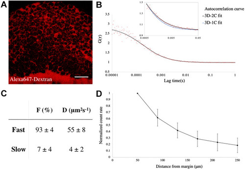
Fgf8a-EGFP propagates by diffusing via the extracellular space. (A) Extracellular space (ECS) in an early gastrula embryo, as visualized by Alexa-647-tagged Dextran injection. The orientation of the embryo is as in Fig. 2A. Scale bar: 50 μm. (B) FCS autocorrelation curve from the ECS of a Tg(fgf8a:fgf8a-EGFP) embryo fitted with 2-C (black curve) and 1-C (blue curve in inset) 3D-diffusion models. (C) Results from fitting with the 3D-2C diffusion model. F, fraction of each component; D, diffusion coefficient. n=52 measurements. (D) Plot of count rate, derived from FCS analysis versus distance from the margin at early gastrula showing graded distribution of Fgf8a in the ECS along the animal-vegetal axis. N=20 embryos. Data are mean±s.d.
|