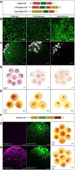
Extracellular diffusion of Fgf8a is necessary for its morphogenic activity. (A) The three different versions of Fgf8a were used in this study. sp, signal peptide; TM, transmembrane. (B) Confocal images of late blastula-stage embryos injected with the various constructs at the one-cell stage to assess localization. Scale bars: 20 μm. (C) Generation of ectopic clones of the constructs and imaging at late blastula stages to determine protein spreading. Co-injection with HRas-mKate labels the source cells (magenta). Scale bars: 50 μm. (D) In situ hybridization against spry4 at late blastula after generating ectopic clones as in C. (E) Double in situ hybridization against gfp (red) and spry4 (blue) in such embryos at late blastula to depict the ectopic source and target gene induction domains, respectively. Scale bars: 100 μm. (F) Schematic of the Morpho-trap construct. (G,H) Late blastula-stage embryos injected at the 32-cell stage with Fgf8a-EGFP (G) or the Morpho-trap at the one-cell stage followed by Fgf8a-EGFP at the 32-cell stage (H). Confocal images are shown in the first two panels. Scale bars: 50 μm. The third panel depicts double in situ hybridization against gfp (red) and spry4 (blue) in such embryos to indicate the Fgf8a-EGFP clone and target gene induction domain, respectively. Scale bars: 100 μm. Animal pole views of embryos are shown throughout.
|
