
Figure 6.
- ID
- ZDB-FIG-230413-12
- Publication
- Goldblatt et al., 2023 - Neuronal birthdate reveals topography in a vestibular brainstem circuit for gaze stabilization
- Other Figures
- All Figure Page
- Back to All Figure Page
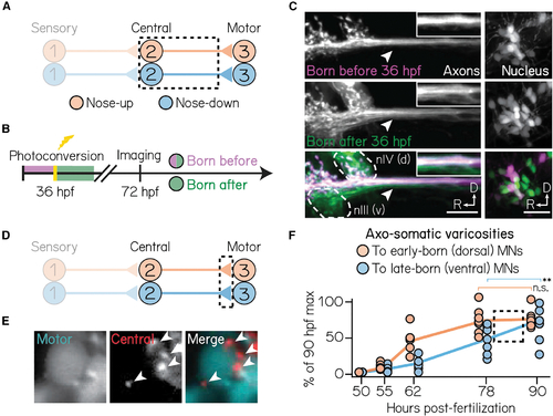
Birthdate predicts patterns of axonal trajectories and synapse formation between projection neurons and extraocular motor neurons
(A) Circuit schematic for axon birthdating experiments. Black dashed lines outline projection neuron soma and axonal projections to the extraocular motor nuclei. (B) Timeline of axon birthdating experiments. Larvae are only photoconverted at 36 hpf. (C) Birthdated axons from one example fish. Top row shows axons (left) from soma (right) born by 36 hpf; middle row shows axons that were not born by 36 hpf; bottom row shows merge. White arrows point to the ventral axon bundle. Inset shows zoom of axons. Dotted lines outline extraocular motor neuron somata. Scale bars, 20 μm. (D) Circuit schematic for synapse formation measurements. Black dashed lines outline axo-somatic varicosities from projection neurons onto post-synaptic ocular motor neurons. (E) Example image of an extraocular motor neuron (left) and axo-somatic varicosities (middle); right image shows merge. White arrows point to four example varicosities. Scale bars, 5 μm. (F) Rate of varicosity growth to early- and late-born ocular motor neurons, quantified as as a percentage of the maximum number of varicosities observed at 90 hpf. Circles represent individual fish. Dotted box highlights the time where the rate of varicosity formation to motor neuron subtypes diverges. n ≥ 5 fish per time point. **difference at the p < 0.01 level; n.s., not significant. |