
Figure 2
- ID
- ZDB-FIG-230217-26
- Publication
- Panlilio et al., 2023 - Developmental exposure to domoic acid targets reticulospinal neurons and leads to aberrant myelination in the spinal cord
- Other Figures
- All Figure Page
- Back to All Figure Page
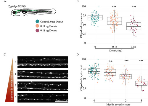
Exposure to DomA at 2 dpf reduces the number of myelinating oligodendrocytes in fish with severe myelin defects in a dose dependent manner. (A) The Tg(mbp:EGFP) transgenic line was used to quantify myelinating oligodendrocytes at 4 dpf. The rectangle delineates the approximate location within the spinal cord where the image was acquired (somites 6–10). (B) Number of myelinating oligodendrocytes quantified at 4 dpf in the spinal cords of fish exposed to different doses of DomA at 2 dpf. Each point represents the number of myelinating oligodendrocytes within the 403.9 μM imaging area in a single fish. (Control 0 ng – median = 70 and IQR = 13; DomA 0.14 ng – median = 62, IQR = 20; DomA 0.18 ng – median = 47, IQR = 20). (C) Representative images of laterally mounted Tg(mbp:EGFP) fish classified by myelin severity, ranging from 0, representing control-like myelin sheaths, to 3, representing the most severe myelin phenotype observed. (D) Fish used in (B) were further subdivided by the severity of the myelin defect observed. The myelinating oligodendrocyte counts were then plotted by the myelin severity score. Scale bar = 100 μm, n.s. = not significant, *** p < 1 e−3 using a generalized mixed effects model with a negative binomial distribution. |