
Fig. 2
- ID
- ZDB-FIG-220923-95
- Publication
- Bensimon-Brito et al., 2021 - Integration of multiple imaging platforms to uncover cardiovascular defects in adult zebrafish
- Other Figures
- All Figure Page
- Back to All Figure Page
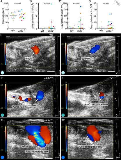
alk5a –/– adult zebrafish display variable haemodynamic defects. (A–D) Parameter quantification obtained with echocardiography analyses of WT (n = 10) and alk5a–/– (n = 12) adult zebrafish, including heart rate (A), area of the aortic flow (B), and regurgitation fraction in the AV (C) and OFT (D) canals. Plots show the values for each individual and the mean ± SD; P-values were determined by unpaired t-test (A) or Mann–Whitney test (B–D). (E, E’) WT zebrafish exhibit unidirectional blood inflow (red, E) and outflow (blue, E’) without signs of regurgitation. (F–G’) Examples of alk5a–/– zebrafish exhibiting a detectable aortic flow (F) and retrograde blood flow (F’–G’). The colour of each dot refers to the same zebrafish across all graphs and images. The dot adjacent to the number symbol (#) identifies the individual zebrafish mentioned in the text. Scale bars: 1 mm (E–G’). |
| Fish: | |
|---|---|
| Observed In: | |
| Stage: | Adult |