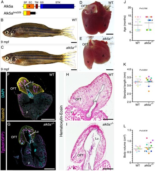
alk5a –/– adult zebrafish display variable cardiac phenotypes without gross morphological defects. (A) Schematic of Alk5a WT and mutant proteins depicting each domain and the site of the mutation (star). (B, C) Brightfield images of 9 mpf WT (B) and alk5a–/– (C) zebrafish. (D, E) Brightfield images of alk5a–/– hearts (E) which occasionally exhibit a dilated outflow tract (OFT) lumen compared to WT (D). (F–I) Cryosections of WT (F, H) and alk5a–/– (G, I) hearts immunostained for Tg(kdrl: eGFP) expression (endothelial cells) and Elastin2 (F, G), and stained for haematoxylin-eosin (H, I) showing the expanded OFT lumen (Lu, dashed line) in alk5a–/– zebrafish. (J–L) Quantification of age (J), standard length (K), and body volume (L) of WT (n = 10) and alk5a–/– (n = 12) zebrafish used in the subsequent analyses. Plots show the values for each individual and the mean ± SD; P-values were determined by unpaired t-test (J, L) or Mann–Whitney test (K). The colour of each dot refers to the same zebrafish across all graphs. The dot adjacent to the number symbol (#) identifies the individual zebrafish mentioned in the text. Scale bars: 2 mm (B, C), 200 µm (D, E, H, I), 400 µm (F, G). A, atrium; EC, extracellular; GS, glycine–serine rich; SP, signal peptide; STK, serine–threonine kinase; TM, transmembrane; V, ventricle.
|
