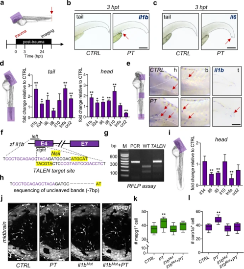
a Experimental setup. hpt, hours post-trauma. b, c The expression of il1b + and il6 + cells (red arrow) in zebrafish larvae was measured by WISH at 3 hpt. Scale bar, 200 µm. d qPCR showed that the mRNA levels of various cytokines or chemokines changed in the traumatic site/tail and the brain/head compared to CTRL. Independent t test, *, p < 0.05, **, p < 0.01. e WISH showed increased il1b + cells (red arrow) in the brain/head, body/trunk, and traumatic site/tail of zebrafish larvae at 24 hpt. Scale bar, 100 µm. h, head. b, body. t, tail. f Design of the transcription activator-like effector nucleases (TALEN) targeting zebrafish il1b. g, h il1b mutation (il1bMut) was confirmed by the restriction fragment length polymorphism (RFLP) assay and Sanger sequencing (−7 bp). (i) qPCR showed that the mRNA levels of various cytokines were decreased in il1bMut zebrafish brains compared to CTRL brains. Independent t test, **, p < 0.01. j–l il1bMut rescued the increased mpeg1 + macrophages or coro1a + leukocytes in the zebrafish brain at 24 hpt. Two-way ANOVA with Tukey’s HSD post hoc test, **, p < 0.01 compared to CTRL. Scale bar, 40 µm.
|
