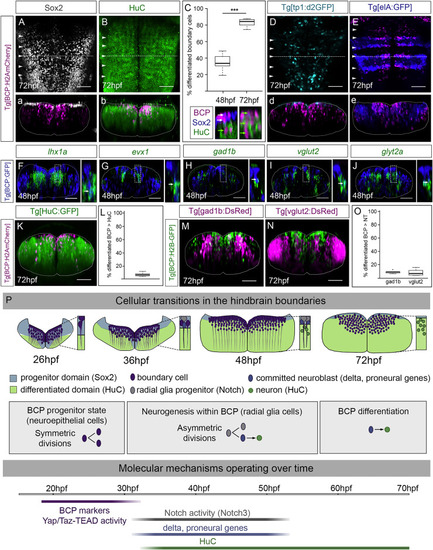
Figure 7. Boundary cells are fated to give rise to neurons (A and B) Tg[BCP:H2AmCherry] embryos immunostained with Sox2 (A) and HuC (B) at 72 hpf. (C) Boxplot displaying the differentiated boundary cells at 48 hpf (34.9%, SD 9.28%, SEM 2.93%) and 72 hpf (82.6%, SD 4.89%, SEM 1.55%; p value paired t test p < 0.001 [∗∗∗]). N = 10 boundaries, n = 5 embryos, two boundaries per embryo. Note magenta cells located in the ventricular zone expressed Sox2 at 48 hpf (white arrows) but not at 72 hpf (HuC cells, green arrows); the displayed region corresponds to the framed area in (b). (D) Tg[BCP:H2AmCherry;tp1:d2GFP] embryos with no boundary progenitor cells displaying Notch-activity at 72 hpf. (E) Tg[BCP:H2AmCherry;elA:GFP] embryos showing rhombomere integrity. (A–B; D–E) Dorsal views with anterior to the top. (a–b; d–e) Boundary transverse views (dashed line). White arrowheads indicate the position of the hindbrain boundaries. (F–J) Tg[BCP:GFP] embryos in situ hybridized with lhx1a (F), evx1 (G), gad1b (H), vglut2 (I), and glyt2a (J) at 48 hpf. Transverse views through r4/r5 or r5/r6; see dorsal views in Figures S6B–S6F. Inserts are magnifications of the corresponding framed regions. Note co-expression of GFP with differentiation factors and neurotransmitters (white arrows in inserts). (K–O) Quantification of the differentiated neurons derived from the boundary cell population. (K, M–N) Tg[BCP:H2AmCherry;HuC:GFP], Tg[BCP:H2B-GFP;gad1b:DsRed], and Tg[BCP:H2B-GFP;vglut2:DsRed] embryos, respectively, as MIP of r4/r5. (L, O) Boxplots with the percentage of differentiated neurons (HuC) (6.5%, n = 7 embryos) and GABAergic and glutamatergic neurons (8.8% GABAergic versus 7.9% glutamatergic; n = 6 embryos each) derived from whole boundaries. BCP, boundary cell population; hpf, hours post-fertilization. Scale bar, 50 μm. (P) Illustration depicting the cellular and functional transitions of the boundary cell population. When neuronal differentiation starts in the hindbrain (26 hpf), boundary cells are progenitors dividing symmetrically (purple). They activate Notch3 signaling (32 hpf), transition to radial glia cells (gray) and undergo asymmetric cell division giving rise to one progenitor (gray) and one committed neuroblast (blue). By 48 hpf, the progenitor domain is reduced; the boundary progenitors still display Notch-activity, whereas the ones committed to the neuroblast lineage differentiate into neurons (green). Finally, the pool of boundary progenitor cells almost extinguishes. Several molecular mechanisms operate over time. Boundary cells are specified at the interfaces between rhombomeres (18–20 hpf) and trigger Yap/Taz-TEAD in response to mechanical stimuli remaining as proliferating progenitors to expand the progenitor pool (Voltes et al., 2019). By 32 hpf, boundary cells are Notch3 active and engage into neurogenesis. Finally, boundary cells most probably undergo direct differentiation, as the differentiation domain dramatically increases at the expense of the progenitor domain.
|
