|
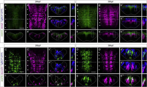
Figure 5. Notch players are expressed in the boundary cells from 36 hpf onward (A–D) Tg[BCP:GFP] embryos, before (26 hpf) and after (36 hpf) the onset of Notch-activity in the boundary cells, were double in situ hybridized with (A–B) notch3 and deltaD and (C–D) ascl1b and neuroD4, followed by GFP staining to detect the boundary cells. (a–a’’’’, b–b’’’’, c–c’’’’, d–d’’’’) Transverse views of the boundary indicated by the dashed line in (A)–(D), displaying staining combinations according to the color-coded labeling. Inserts display magnifications of a single-stack view of the corresponding framed region. Note that notch3 and deltaD staining never overlap. (A–A′, B–B′, C–C′, D–D′) Dorsal MIP with anterior to the top. White arrowheads indicate the position of the hindbrain boundaries. BCP, boundary cell population; hpf, hours post-fertilization. Scale bar, 50 μm.
|