
Figure 7
- ID
- ZDB-FIG-220131-58
- Publication
- Brombin et al., 2022 - Tfap2b specifies an embryonic melanocyte stem cell that retains adult multifate potential
- Other Figures
- All Figure Page
- Back to All Figure Page
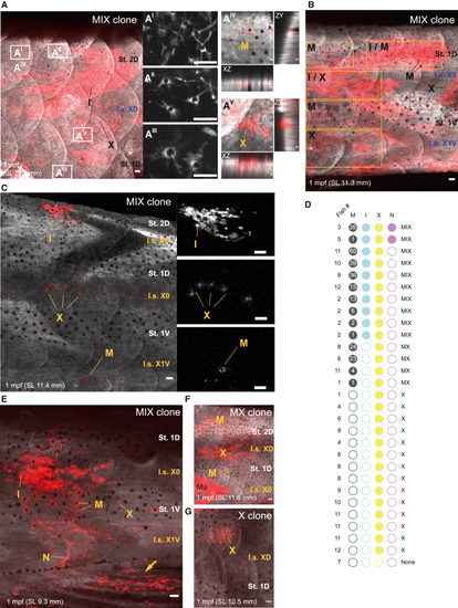
tfap2b+ McSCs have multifate potential for all adult pigment cell lineages (A–C) MIX clones in tfap2b lineage tracing analysis in the adult pigment pattern. Shown are the (A) caudal trunk, (B) tail region, and (C) medial trunk. Magnified images of melanocytes are presented in stripes 2D (AI and AII) and 1D (AIII). Orthogonal projections of a melanocyte (AIV) and a xanthophore (AV) show that cells are localized in the scale. Also shown are (B) a MIX clone spanning the dorsoventral axis and (C) a sparse MIX clone, with magnifications of iridophores, xanthophores, and a melanocyte (top to bottom). White pseudocoloring is used for the GFP channel, and red is used for the mCherry channel in (A)–(C). white pseudocoloring is used for the mCherry channel in the magnified panels and AI–AIII and in (C) magnified images. Shown are representative images of more than 20 fish injected with 25 pg/nL of tfap2b:cre. MAX projection. Scale bars, 50 μm. (D) Frequencies of the different derivatives in clearly defined clones in 12 juvenile ubi:switch zebrafish injected with low doses of the tfap2b:cre plasmid (1.5 pg/nL, 3.25 pg/nL, and 6 pg/nL). Fish SL in millimeters: (1) 9.7, (2) 8.7, (3) 7.1, (4) 10.5, (5) 9.3, (6) 11.6, (7) 11.1, (8) 10.6, (9) 12.4, (10) 11.2, (11) 13.7, and (12)15.5. Clones were named according to their pigment cell composition. Numbers in the black dots represent mCherry+ melanocytes within the clone. (E−G) Representative images of the clones analyzed in (D). Shown are (E) a MIX clone with a clone-associated nerve (caudal trunk; the arrow indicates clone extension into the anal fin), (F) an MX clone (medial trunk), and (G) an X clone (rostral trunk). MAX projection. Scale bars, 50 μm. All fish were injected with 1.5 pg/nL of tfap2b:cre. I.s., interstripe; St., stripe. See also Figure S6. |