|
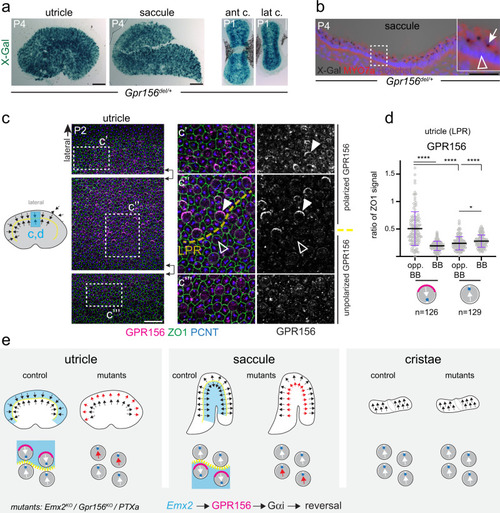
<italic>Gpr156</italic> expression and GPR156 protein localization in mouse vestibular organs.aLacZ reporter is expressed throughout the sensory region in Gpr156del/+ vestibular organs (ant/lat c., anterior/lateral crista). bLacZ expression is limited to MYO7A+ HCs in a saccule cross-section. X-gal signal is trapped in HC vesicles (arrow in magnified inset) but support cells (arrowhead) are negative. c, d P2 wild-type utricle where basal body labeling (PCNT) indicates HC orientation. GPR156 polarization (solid arrowheads) is limited to lateral HCs oriented medially. HCs across the LPR oriented laterally do not show polarized GPR156 (hollow arrowheads). Boxed regions in continuous fields in the left panels are magnified in the central and right panels (saccule: see Supplementary Fig. 2c). d GPR156 enrichment in the utricle LPR domain at the HC junction opposite (opp. BB) or near (BB) the basal body. HCs oriented medially (left) are analyzed separately from HC oriented laterally (right). GPR156 is expressed as ratio of ZO1 signal (mean ± SD; n, HC numbers in 3 animals; Kruskal-Wallis test with Dunn’s multiple comparisons, ****p < 0.0001; *p = 0.0332). e Summary of HC orientation (arrows), GPR156 protein distribution (magenta) and previously reported Emx2 expression (blue) by vestibular organ in normal and mutant conditions. The scheme in c indicates the position of the domain analyzed in c and d (blue). Scale bars are 100 µm (a), 50 µm (b), 20 µm (c).
|