|
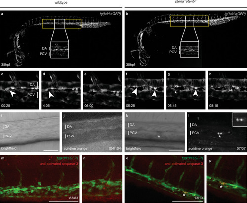
A population of HSPCs fails to complete EHT and undergoes apoptosis in <italic>ptena</italic><sup><italic>−/−</italic></sup><italic>ptenb</italic><sup><italic>−/−</italic></sup> mutant embryos.a, b Brightfield image of a wild-type or ptena−/−ptenb−/− mutant zebrafish embryo at 35 hpf. The area from which HSPCs originate is indicated with a yellow box. A close up is indicated with a white box. c–h Four-dimensional imaging of tg(kdrl:eGFP) wild-type or ptena−/−ptenb−/− mutant embryos between 35 and 48 hpf. Still frames from Movie S1. Arrowheads: HSPCs undergoing EHT; asterisk: disintegrating HSPCs. Confocal image z-stacks (2 µm step size, with ×40 objective and ×2 zoom; anterior to the left; maximum projections of a representative embryo; time in hh: mm. i–l Acridine orange staining. Arrows and circles: HSPCs in VDA of 40–45 hpf embryos. Asterisks: apoptotic HSPCs. Scale bar: 50 µm. Representative embryos are shown and the number of embryos that showed this pattern/total number of embryos is indicated. DA dorsal aorta, PCV posterior cardinal vein. m–p confocal images of apoptotic endothelial cells in the VDA of fixated wild-type or ptena−/−ptenb−/− mutant zebrafish embryos. In green: tg(kdrl:eGFP); in red: anti-activated caspase-3 immunohistochemistry staining. Apoptotic cells are indicated with an asterisk. Representative embryos are shown and the number of embryos displaying this particular pattern/total number of embryos is indicated in the bottom right. Anterior to the left; 2 µm step size; maximum projections; scale bar: 100 µm.
|