
Fig. 4
- ID
- ZDB-FIG-210210-7
- Publication
- Cambier et al., 2020 - Spreading of a mycobacterial cell surface lipid into host epithelial membranes promotes infectivity
- Other Figures
- All Figure Page
- Back to All Figure Page
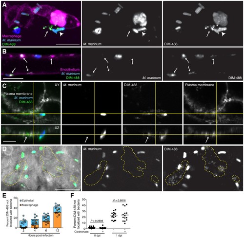
PDIM spreads into epithelial membranes. (A) Image of M. marinum expressing a cytosolic blue-fluorescent protein recoated with DIBO-488 labeled azido-DIM (DIM-488) highlighting DIM-488 spread from bacteria (arrowhead) to epithelial cells (arrows) at 3 hpi of ~100 M. marinum in the HBV, scale bar = 10 μm. (B) Image of DIM-488 labeled M. marinum at 1 day post-intravenous infection of transgenic fish whose endothelium express a red-fluorescent protein. Arrows, DIM-488 spread onto endothelium, scale bar = 5 μm. (C) Image of A549 epithelial cells whose plasma membranes are labeled with Alexa-fluor 594 wheat germ agglutinin at one day post infection with DIM-488 labeled M. marinum at an MOI of 5. Arrows, DIM-488 spread into plasma membrane, scale bar = 5 μm. (D) Image highlighting DIM-488 spread onto epithelial surfaces (yellow-dashed outline) at 2 hpi of ~100 M. marinum in the HBV, scale bar = 10 μm. (E) Mean percent DIM-488 in macrophage or epithelial cells not localized with bacteria following HBV infection with ~100 M. marinum. Representative of two separate experiments. (F) Mean percent DIM-488 not localized with bacteria following HBV infection of lipo-PBS or lipo-clodronate treated fish with ~100 M. marinum. Two-tailed Mann Whitney test for 0 dpi and two-tailed, unpaired t test for one dpi. Representative of three separate experiments. |