Figure 1; supplement 3.

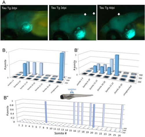
Movement of some Tau puncta over time following injection of zebrafish larvae with brain homogenate burdened with human tauopathy.

(A) Images of the zebrafish brain area, after injection with Tg hTau+/p301L mouse brain homogenate, for the same zebrafish larvae over 3 consecutive days post-injection (dpi), showing the movement of one puncta over time. (B, B’ and B”) The location of Tau aggregates on the spinal cord of three exemplar individual larvae over multiple days, denote movement of some of these puncta over time. Somite numbers were used as landmarks to report the location along the spinal cord over multiple days - somites are blocks of tissue (e.g. muscle) that are readily visible in the trunk of the larva; larger somite number are more posterior (i.e. more distal to the injected brain region) with the most distal region being ‘unsegmented’.

Expression Data

Expression Detail
Antibody Labeling
Phenotype Data

Phenotype Detail
Acknowledgments
This image is the copyrighted work of the attributed author or publisher, and ZFIN has permission only to display this image to its users. Additional permissions should be obtained from the applicable author or publisher of the image. Full text @ Elife