Figure 1; supplement 2.

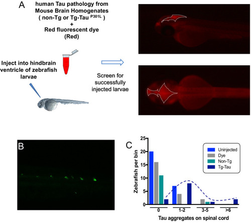
Quantification GFP+ inclusions in CNS of zebrafish expressing Tau4R-GFP biosensor, following injection of human Tau into the zebrafish hindbrain ventricle.

(A) Schematic showing the microinjections of mouse brain homogenate into the brain ventricles of zebrafish larvae (age is 2 days post-fertilization). Injected material was brain homogenate from transgenic mice expressing human Tau, that is with Tau pathology (e.g. see pathology characterized in Eskandari-Sedighi et al., 2017, DOI 10.1186/s13024-017-0215-7) or brain homogenate from normal control mice. Red fluorescent dye included in the diluted brain homogenate allowed imaging and selection of properly injected embryos. Right: Successfully injected embryos had the injected brain homogenate localized within the zebrafish brain ventricles, with sharp edges and non-diffuse dye. The area encompassing the injected dye is outlined by white dotted lines. (B) Similar to Tau4R-GFP aggregating in the brain, GFP+ aggregates were also detected in the spinal cord following delivery of human Tau, akin to the brain aggregates in Figure 1F. (C) Quantification of spinal cord aggregates. Same data as Figure 1G re-plotted to show the distribution of larvae displaying various quantities of aggregates in the spinal cord. Color coding of treatments equivalent to Figure 1G. Dotted line is hand drawn to emphasize the distribution of larvae injected with tau-burdened brains, compared to various the controls with peak distributions of GFP+ puncta near to zero.

Expression Data

Expression Detail
Antibody Labeling
Phenotype Data

Phenotype Detail
Acknowledgments
This image is the copyrighted work of the attributed author or publisher, and ZFIN has permission only to display this image to its users. Additional permissions should be obtained from the applicable author or publisher of the image. Full text @ Elife