Figure 1; supplement 1.

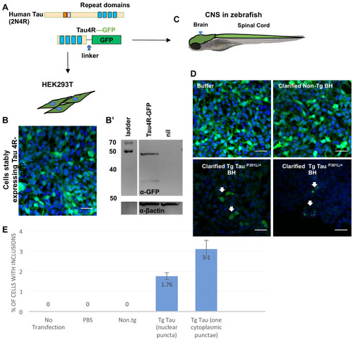
Quantification GFP+ inclusions in HEK cells expressing Tau4R-GFP biosensor.

(A) Schematic of genetically encoded fluorescent Tau4R-GFP ‘Tau Biosensor’ which was produced by fusing the C-terminal four binding repeats (4R) region of wild-type human Tau (Tau4R) to GFP via a linker. This construct was expressed as a transgene (C) in the zebrafish CNS under the promoter enolase 2, and (B) in human embryonic kidney HEK293T cells for validation. (B’) Immunoblot vs. GFP suggests the fusion protein Tau4R-GFP remains intact as a fusion protein when expressed in HEK cells and is an appropriate size (similar to when expressed in zebrafish CNS, Figure 1C). (D) Tau4R-GFP biosensor cells display GFP+ inclusions only upon transduction of crude brain homogenate burdened with tauopathy (from Tg mice expressing human TauP301L), but not when transduced with brain homogenate from normal mice (non-Tg BH). (E) HEK cells from D had a detection rate of approximately 2–3% of cells having GFP+ puncta, whereas various negative controls (no transfection, transfection with PBS only, or transfection of brain homogenate from non-Tg control mice) consistently showed no puncta. For the quantification, the number of cells with inclusions and the number of total cells from nine field images were counted. Bars represents mean of three independent experiments.

Expression Data

Expression Detail
Antibody Labeling
Phenotype Data

Phenotype Detail
Acknowledgments
This image is the copyrighted work of the attributed author or publisher, and ZFIN has permission only to display this image to its users. Additional permissions should be obtained from the applicable author or publisher of the image. Full text @ Elife