FIGURE

Figure 8

ID
ZDB-FIG-201107-26
Publication
Costa et al., 2020 - RAB13 mRNA compartmentalisation spatially orients tissue morphogenesis
Other Figures
All Figure Page
Back to All Figure Page
Figure 8

<italic>rab13</italic> mRNA polarisation orients blood vessel morphogenesis

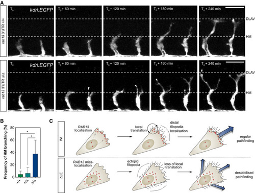
Time‐lapse confocal microscopy of representative Tg(kdrl:EGFP) rab13+/+ and rab13∆3′UTR/∆3′UTR embryos. DLAV: dorsal longitudinal anastomotic vessel; HM: horizontal myoseptum.

Frequency of ISV ectopic branching occurring at the HM (= 4 experiments; *< 0.05; one‐way ANOVA with Bonferroni's correction).

Illustration of the role for RAB13 mRNA localisation, local translation and compartmentalisation of RAB13 function in defining the orientation of EC filopodia dynamics, motile EC polarity and blood vessel pathfinding.

Data information: T0 = 25 hpf; arrowheads indicate extra branches emerging from the main ISVs at the HM position; scale bars = 50 μm (A). +/+, +/∆ and ∆/∆ represent Tg(kdrl:EGFP) rab13+/+, rab13+/∆3′UTR and rab13∆3′UTR/∆3′UTR embryos, respectively (B). Bar chart is presented as means ± s.d.

Expression Data

Expression Detail
Antibody Labeling
Phenotype Data
Fish:
Observed In:
Stage: Prim-5

Phenotype Detail
Acknowledgments
This image is the copyrighted work of the attributed author or publisher, and ZFIN has permission only to display this image to its users. Additional permissions should be obtained from the applicable author or publisher of the image. Full text @ EMBO J.