FIGURE

Figure EV5

ID
ZDB-FIG-201107-24
Publication
Costa et al., 2020 - RAB13 mRNA compartmentalisation spatially orients tissue morphogenesis
Other Figures
All Figure Page
Back to All Figure Page
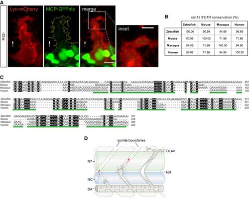
Figure EV5

The 3′<styled-content toggle='no' style='fixed-case'>UTR</styled-content> of <italic>rab13</italic> shows low conservation across species whilst retaining <styled-content toggle='no' style='fixed-case'>mRNA</styled-content> localisation potential

Tg(fli1ep:MCP‐GFPnls) tip cell expressing a control Lyn‐mCherry‐24xMS2 construct.

Percentage identity matrix of rab13 3′UTR orthologue sequences.

Multiple sequence alignment between rab13 3′UTR orthologues. Black boxes indicate absolute nucleotide similarity. The human RAB13 3′UTR localisation element is underlined in green.

Scheme depicts stages of zebrafish ISV sprouting. DA: dorsal aorta; DLAV: dorsal longitudinal anastomotic vessel; HM: horizontal myoseptum; NC: notochord; NT: neural tube.

Data information: white arrows indicate direction of ISV sprouting; yellow dashed line outlines ISV cell borders; scale bars = 10 μm; scale bar in inset = 5 μm (A).

Expression Data

Expression Detail
Antibody Labeling
Phenotype Data

Phenotype Detail
Acknowledgments
This image is the copyrighted work of the attributed author or publisher, and ZFIN has permission only to display this image to its users. Additional permissions should be obtained from the applicable author or publisher of the image. Full text @ EMBO J.