|
Figure 8
Time‐lapse confocal microscopy of representative Frequency of ISV ectopic branching occurring at the HM ( Illustration of the role for
Data information:

|
|
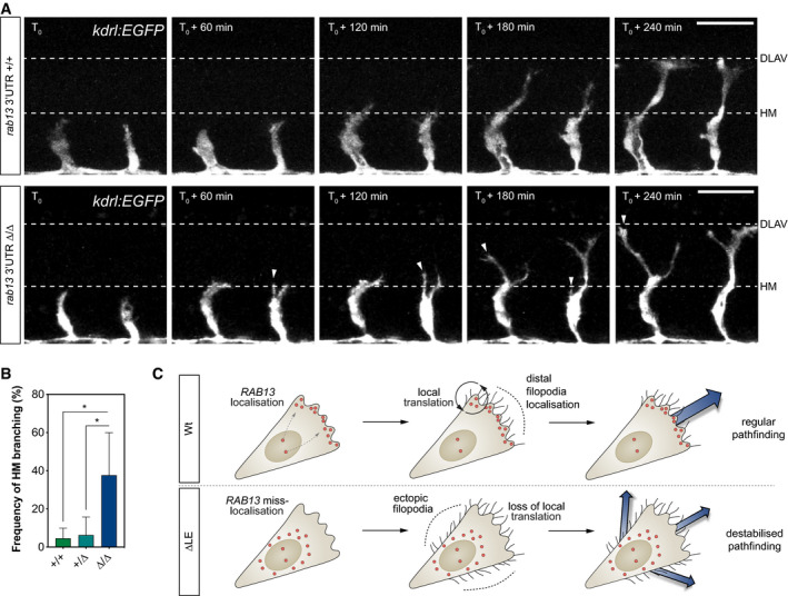
Figure 8
Time‐lapse confocal microscopy of representative Frequency of ISV ectopic branching occurring at the HM ( Illustration of the role for
Data information: