|
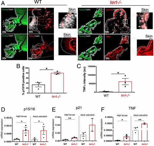
tert −/− tissues present increased levels of senescence (p15/16) and inflammation (TNF-α). (A) Representative immunofluorescence images of p15/16 and TNF-α in the skin near melanoma in chimeric WT and tert−/− zebrafish. Dashed lines locate the skin (no green fluorescence), and arrows indicate p15/16-positive cells. Squares with dashed lines show place of amplification in a sequential section, and squares with solid lines show place of amplification on that section. (B and C) Quantification of p15/16-positive cells and levels of TNF-α, respectively, in the skin of chimeric WT and tert−/− zebrafish (n = 3). (D–F) RT-qPCR analysis comparing the expression levels of cdkn2a/b (p15/16), cdkn1a (p21), and tnfa (TNF) of 4-dpf WT, G2 tert−/− larvae, 9-months WT, and tert−/− adult intestine tissue. Data are represented as mean ± SEM. (*P < 0.05; **P < 0.01 for n = 30).
|