FIGURE

Fig. 1

ID
ZDB-FIG-200730-8
Publication
Hall et al., 2020 - In vivo cell biological screening identifies an endocytic capture mechanism for T-tubule formation
Other Figures
All Figure Page
Back to All Figure Page
Fig. 1

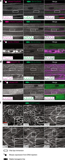
An in vivo system for T-tubule development.

ae The CaaX domain from Hras marks both sarcolemma and T-tubule. Markers were screened at 48 hpf for T-tubule specificity on a stable transgenic background expressing Bin1b-Citrine from the endogenous locus. a CaaX domain from Hras (tH). b CaaX domain from Kras (tK). c Myristoylation domain from ARF5. d BODIPY-TR vital dye. e GPI anchor from zebrafish folate receptor gamma. f Time course of tubule formation using stable EGFP-CaaX background. All images are representative of 12 individual cells within different individual animals. See also Supplementary Movies 1 and 2. Scale bars; (ae) 10 µm, (f) 50 μm.

Expression Data
Genes:
Fish:
Anatomical Terms:
Stage Range: 14-19 somites to Long-pec

Expression Detail
Antibody Labeling
Phenotype Data

Phenotype Detail
Acknowledgments
This image is the copyrighted work of the attributed author or publisher, and ZFIN has permission only to display this image to its users. Additional permissions should be obtained from the applicable author or publisher of the image. Full text @ Nat. Commun.