|
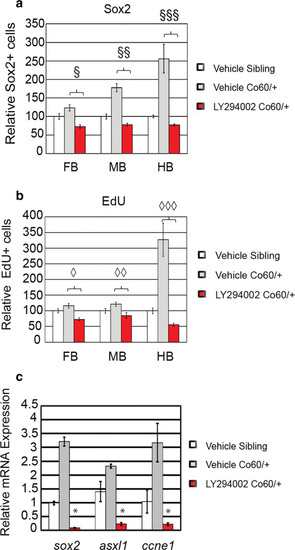
Quantification of the number of NPCs after inhibition of Asxl1 activity. a, b Quantification of the number of Sox2+ (a) or EdU (b) positive cells in vehicle treated and larvae treated with LY294002 at 2 days post fertilization (DFP). N = 4 Vehicle Sibling, 6 Vehicle hcfc1aco60/+ larvae, and 4 LY294002 hcfc1aco60/+ larvae. §p = 0.000168, §§p = 2.08852E−09, §§§p = 0.000153. ◊p = 7.22142E−07, ◊◊p = 0.000997, ◊◊◊p = 0.00014. All error bars represent standard error of the mean. chcfc1aco60/+ larvae and their wildtype siblings (Sibling) were treated at 24 h intervals with 12 μM LY294002 until 5 DPF and then total RNA was isolated from brain homogenates. Quantitative real time PCR was performed to test the expression of sox2, asxl1, and cyclin E (ccne1). N = 10/group/biological replicate. Error bars represent standard error of the mean. *p < 0.05
|