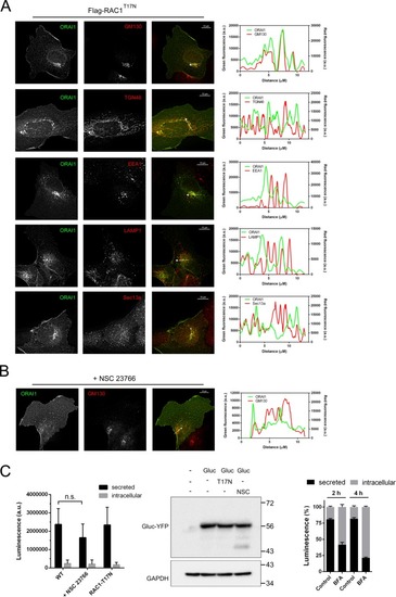
RAC1T17N blocked the transport of ORAI1 to the cell surface. Panel A: U2OS cells stably expressing Flag-RAC1T17N were transfected for the transient expression of ORAI1-GFP. Cells were fixed 24 h after transfection, and used for the immunolocalization of GM130, TGN46, EEA1, LAMP1, and Sec13a. Secondary antibodies were labelled with Alexa Fluor 594. Images are representative of 2 independent experiments (>20 cells per condition). Fluorescence of both channels was measured over the arrow depicted in the image, which was placed over the intracellularly trapped GFP- signal. Panel B: U2OS cells were treated with 50 μM NSC 23766 for 8 h, fixed, and the immunolocalization of GM130 was performed as in panel A. Images are representative of 26 cells from 2 independent experiments. Bar = 10 μm. Panel C: Left: Cells were cultured on 96-well plates in DMEM + 10% FBS medium. The secreted and the intracellular luciferase activity were measured after 28 h of culture. The treatment with 50 μM NSC 23766 was performed during the last 8 h of culture. The overexpression of Flag-RAC1T17N was triggered with doxycycline during the last 22 h of culture. Data from n = 15 wells and 3 independent experiments are shown as bar chart. Middle: Intracellular Gluc-YFP protein was evaluated from cell lysates by immunoblot using an anti-GFP antibody. Right: Cells were treated with 5 μg/ml brefeldin A for 2–4 h to assess the inhibition of the secretory pathway, as a control of the experiment.
|
