FIGURE

Figure 3

ID
ZDB-FIG-200427-7
Publication
Lopez-Guerrero et al., 2020 - RAC1-Dependent ORAI1 Translocation to the Leading Edge Supports Lamellipodia Formation and Directional Persistence
Other Figures
All Figure Page
Back to All Figure Page
Figure 3

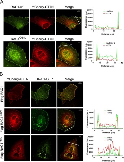
Activation of RAC1 triggered the translocation of ORAI1 to CTTN-containing cell periphery. Panel A: Cells growing onto collagen-coated coverslips in FBS-containing DMEM were transfected for the expression of mCherry-CTTN and EGFP-RAC1 (wt) or EGFP-RAC1Q61L (active RAC1). At 36 h after transfection, cells were fixed and visualized under wide-field epifluorescence microscopy. Cherry and GFP channels were recorded sequentially using independent filter blocks. The two images were merged, and the fluorescence of both channels was measured over the arrow depicted in the image. Fluorescence values, plotted on right panels, were quantified with the NIS-Elements AR software. Images are representative of a minimum of 32 cells per condition from 3 independent experiments. Bar = 10 μm. Panel B: Cells stably expressing Flag-RAC1 (wild-type), Flag-RAC1G12V, or Flag-RAC1T17N were transfected for the expression of mCherry-CTTN and ORAI1-GFP. As in panel A, cells were analyzed under epifluorescence microscopy, and Cherry and GFP channels were recorded. Images are representative of >26 cells per condition from 3 independent experiments. Bar = 10 μm.

Expression Data

Expression Detail
Antibody Labeling
Phenotype Data

Phenotype Detail
Acknowledgments
This image is the copyrighted work of the attributed author or publisher, and ZFIN has permission only to display this image to its users. Additional permissions should be obtained from the applicable author or publisher of the image. Full text @ Sci. Rep.