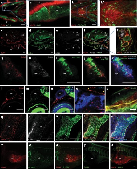
Spatio-temporal pattern of ColVI distribution in larvae from wild-type animals and transgenic reporter fish lines. (a-a’) Individual z-stack of whole-mount immunofluorescence for ColVI (red) and collagen II (green) in 3-dpf larvae. Ventral view. A strong ColVI labeling in evident in the connective tissue surrounding craniofacial cartilages and in the jaw joint. Panel a’ is a magnification of the dotted area of panel a, showing the deposition of ColVI in the jaw joint. (b-b’) Confocal z-stacks of whole-mount immunofluorescence for ColVI (red) in 6-dpf Tg(7xTCF-Xla.Siam:GFP)ia4 (TCF-GFP, green) larvae, showing ColVI labeling near Wnt-positive cells in craniofacial cartilages. Ventral view. Panel b’ is a magnification of the dotted area of panel b, showing ColVI labeling in close apposition to Wnt-positive cells in the ceratohyal cartilages. (c-f’) Section of 7-dpf larvae stained with anti-α1(VI) antibodies (red), WGA (green) and anti-MF20 antibodies (gray). ColVI labeling is present in craniofacial cartilaginous elements, as well as in blood vessels (arrowhead). Panel f’ is a magnification of the dotted area of panel f, showing ColVI labeling in the perichondrium surrounding the cartilaginous elements of the pharyngeal arches, as revealed by co-localization with WGA. (g–k) Individual z-stack of whole mount immunostaining of 6-dpf Tg(osx:nuGFP) (osx:GFP, green) larvae operculum, stained with ColVI (red) and collagen XII (gray) antibodies and Hoechst (blue). ColVI and collagen XII surround operculum osteoblasts. (l–n) Section of a 7-dpf larva stained with anti-ColVI antibodies (red), WGA (green) and Hoechst (blue). ColVI labeling is present in the connective tissue around brain, olfactory pit and eye. (o) Section of a 7-dpf larva stained with anti-ColVI antibodies (red) and Hoechst (blue), showing ColVI labeling in the meninges (arrowhead). (p) Confocal z-stacks of whole-mount immunofluorescence for ColVI (red) and collagen XII (green) on 6 dpf larvae eye, showing ColVI deposition in the connective tissue surrounding the eye. Dorsal view. (q–u) Individual z-stack of whole mount immunostaining for ColVI (red), collagen XII (gray) antibodies and Hoechst (blue) in 2-dpf Tg(fli1:EGFP) (fli:EGFP, green) larvae at the level of pharyngeal arches. ColVI and collagen XII are part of the bordering tissue of migrating pharyngeal pouches. (v–x) Confocal z-stacks of whole-mount immunofluorescence for ColVI (red) in 6-dpf Tg(fli1:EGFP) (fli:EGFP, green) larvae. A strong pattern of ColVI labeling is found around blood vessels (arrowheads) of the intestine. (y-z) Confocal z-stacks of whole-mount immunofluorescence for ColVI (red) and collagen XII (green) in 6-dpf larvae, showing that ColVI but not collagen XII is deposited in the intestine. Scale bar, 100 µm (v–x) or 50 µm (a–u,y,z). cb, ceratobranchial; ch, ceratohyal; ey, eye; f, forebrain; h, hindbrain; jj, jaw joint; mc, Meckel’s cartilage; mid, midbrain; op, olfactory pit; ope, operculum; pa, pharyngeal arch; pq, palatoquadrate.
|
