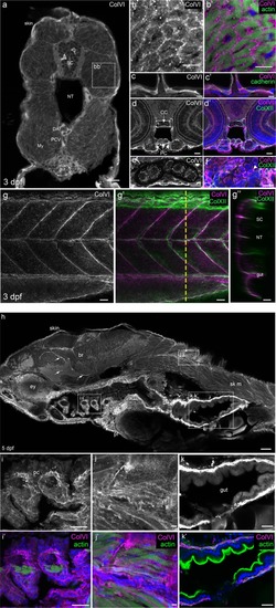
Characterization of ColVI deposition in 3- and 5-dpf developing zebrafish. (a-g’) Immunofluorescence images of transverse sections (a–f) or whole-mount (g-g”) 3-dpf larvae at the level of the trunk. Samples were labeled with anti-ColVI antibodies (panels a-e, g in grey; panels b’,c’,d’,f,g’,g” in magenta) and, where indicated, with phalloidin to reveal actin (green; panel b’), or with antibodies against the epithelial marker cadherin (green; panel c’) and the mesenchymal marker collagen XII (green; panels d’,f,g’,g”). Nuclei were stained with Hoechst (blue; panels b’,c’,d’,f). (a) Global view of ColVI immunoreactivity at the level of the trunk. (b,b’) Zoomed images of the boxed region in (a), showing myotomal muscle cells. (c,c’) Epidermis. (d,d’) Transverse section of the head. (e,f) Zoomed images of the boxed regions of panels d and d’, showing chondrocyte stacks in the cranial cartilage. White arrowhead points to intracellular ColVI staining in chondrocytes. (g) Image of vertical myosepta, obtained from a z-projection of confocal stack of lateral views. (g’) Merge image of the same acquisition, showing ColVI and collagen XII double staining. (g”) Orthogonal view at the level indicated by the yellow dashed line in g’. (h-k’) Immunofluorescence images of sagittal sections of 5-dpf larvae. Samples were labeled with α1(VI) antibodies (grey or magenta) and, where indicated, with phalloidin (green; panels i’,j’,k’). Nuclei were stained with Hoechst (blue; panels i’,j’,k’). (h) Global view of ColVI immunoreactivity. Arrows indicate ColVI-positive vessels. (i-k’) Zoomed images of the boxed regions of panel h. (i,i’) Ceratobranchial cartilage. (j,j’) Skeletal muscle at the trunk level. (k,k’) Intestine. Scale bars, 50 μm (h); 25 μm (d,d’,g-g”,i,i’,k,k’); 20 μm (a); 10 μm (b,b’,e,f); 5 μm (c,c’,j, j’). br, brain; CC, chondrocranium; DA, dorsal aorta; ey, eye; li, liver; My, myotome; NT, notochord; pc, pharyngeal cartilage; PCV, posterior cardinal vein; PC, pharyngeal cartilage; pf, pectoral fin; SC, spinal cord; sk m, skeletal muscle.
|
