
FIGURE
Figure 6
- ID
- ZDB-FIG-200102-6
- Publication
- Tonelotto et al., 2019 - Spatio-temporal expression and distribution of collagen VI during zebrafish development
- Other Figures
- All Figure Page
- Back to All Figure Page
Figure 6
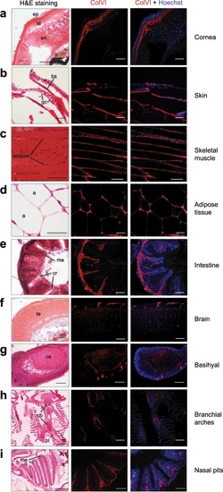
Characterization of ColVI deposition in adult zebrafish. ( |
Expression Data
| Antibody: | |
|---|---|
| Fish: | |
| Anatomical Terms: | |
| Stage: | Adult |
Expression Detail
Antibody Labeling
Phenotype Data
Phenotype Detail
Acknowledgments
This image is the copyrighted work of the attributed author or publisher, and
ZFIN has permission only to display this image to its users.
Additional permissions should be obtained from the applicable author or publisher of the image.
Full text @ Sci. Rep.