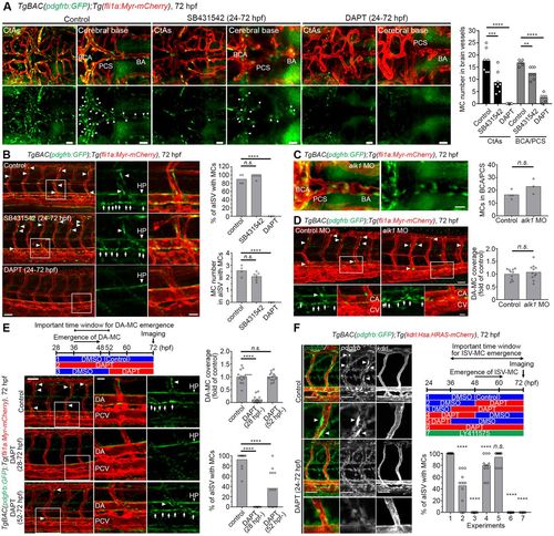
The emergence of pdgfrbhigh MCs was inhibited by DAPT treatment during the specification period. (A,B) Confocal stack images of pdgfrbhigh MCs at 72 hpf in brain vessels (A) or trunk vessels (B) after treatment with vehicle-DMSO (control), 50 µM SB431542 or 100 μM DAPT from 24 hpf through 72 hpf. E3 medium-containing inhibitors were replaced with fresh ones every 12 h. (C,D) Brain vessels (C) and trunk vessels (D) of 72 hpf TgBAC(pdgfrb:GFP);Tg(fli1a:Myr-mCherry) larvae injected with 6 ng alk1 MO. Note that alk1 MO induced abnormal vasculature in the cerebral base or caudal vein but did not inhibit the emergence of pdgfrbhigh MCs. (E) Trunk vessels of TgBAC(pdgfrb:GFP);Tg(fli1a:Myr-mCherry) larvae treated with vehicle-DMSO or 100 µM DAPT at the time point indicated in the top schematic. Boxed regions are enlarged to the right. (F) Confocal stack images of TgBAC(pdgfrb:GFP);Tg(kdrl:Hsa.HRAS-mCherry) larvae treated with either 1% DMSO, 25 μM DAPT or 5 μM LY411575 at the time point indicated in the schematic on the right. Arrows and arrowheads indicate pdgfrbhigh MCs beneath DA and in BSA/PCS/BA/ISVs, respectively. Quantitative results are shown on the right of each representative image. Bars and circles in the graph indicate averages and each value of observed larvae, respectively. ****P<0.0001, ***P<0.001 and **P<0.01, significant difference between control and indicated group. BA, basilar artery; CA, caudal artery; CV, caudal vein; HP, hypochord; n.s., not significant; PCS, posterior communicating segment. Scale bars: 30 µm in A,C; 50 µm in B,D,E; 20 µm in enlarged images in B,D,E; 15 µm in F.