
Fig. 1
- ID
- ZDB-FIG-191007-15
- Publication
- Nicholas et al., 2019 - Temporal characterization of optic fissure basement membrane composition suggests nidogen may be an initial target of remodeling
- Other Figures
- All Figure Page
- Back to All Figure Page
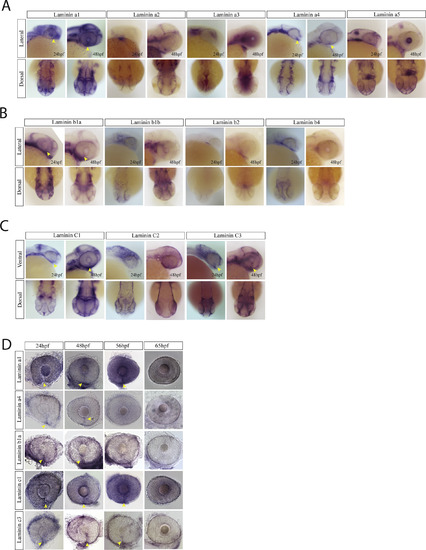
Laminin expression during optic fissure formation and fusion. A) WISH for zebrafish laminin alpha chain genes a1, a2, a3, a4 and a5. Expression was analyzed at 24 and 48hpf and visualized in lateral and dorsal views. At 24 and 48hpf laminins a1, a4 and a5 were observed to express within the optic fissure (yellow arrow heads). Laminin a2 and a3 were largely absent from the retina while laminin a5 was expressed in the developing lens at 48hpf. B) WISH for laminin beta chain genes b1, b2 and b3 at 24 and 48hpf. Only laminin 1b was found to be expressed in the optic fissure (yellow arrow heads). Laminin b2 and b3 were largely absent from the retina. C) WISH for laminin genes c1, c2 and c3 at 24 and 48hpf. Laminin c1 and c3 were detected in the optic fissure (yellow arrow heads) while c2 appears to primarily be expressed in the ectoderm. D) WISH time course examination of laminin gene expression in the optic fissure. Laminin a4 and b1 are expressed in the fissure up to 48hpf, while a1, c1 and c3 retain expression up to 56hpf (yellow arrow heads). |
| Genes: | |
|---|---|
| Fish: | |
| Anatomical Terms: | |
| Stage Range: | Prim-5 to Pec-fin |
Reprinted from Developmental Biology, 452(1), Nicholas, C., Weaver, M., Piedade, W.P., Vocking, O., Famulski, J.K., Temporal characterization of optic fissure basement membrane composition suggests nidogen may be an initial target of remodeling, 43-54, Copyright (2019) with permission from Elsevier. Full text @ Dev. Biol.