
FIGURE
Fig. 2
- ID
- ZDB-FIG-191007-14
- Publication
- Nicholas et al., 2019 - Temporal characterization of optic fissure basement membrane composition suggests nidogen may be an initial target of remodeling
- Other Figures
- All Figure Page
- Back to All Figure Page
Fig. 2
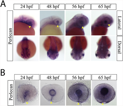
Perlecan expression during optic fissure formation and fusion. A) WISH for zebrafish perlecan at 24, 48, 56 and 65hpf. Perlecan expression is observed in the lens and the optic fissure from 24 to 65 hpf (yellow arrow heads). B) Examining WISH in isolated zebrafish eyes at 24, 48, 56 and 65hpf indicates that perlecan expression persists in the optic fissure (yellow arrow heads) up to 65hpf. |
Expression Data
| Gene: | |
|---|---|
| Fish: | |
| Anatomical Terms: | |
| Stage Range: | Prim-5 to Pec-fin |
Expression Detail
Antibody Labeling
Phenotype Data
Phenotype Detail
Acknowledgments
This image is the copyrighted work of the attributed author or publisher, and
ZFIN has permission only to display this image to its users.
Additional permissions should be obtained from the applicable author or publisher of the image.
Reprinted from Developmental Biology, 452(1), Nicholas, C., Weaver, M., Piedade, W.P., Vocking, O., Famulski, J.K., Temporal characterization of optic fissure basement membrane composition suggests nidogen may be an initial target of remodeling, 43-54, Copyright (2019) with permission from Elsevier. Full text @ Dev. Biol.