
Fig. 6
- ID
- ZDB-FIG-180622-19
- Publication
- Di Donato et al., 2018 - An Attractive Reelin Gradient Establishes Synaptic Lamination in the Vertebrate Visual System
- Other Figures
- All Figure Page
- Back to All Figure Page
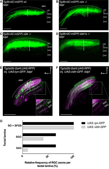
The Reelin Gradient Is a Chemoattractant for Retinal Afferents during Laminar Targeting (A) Side view of the retinotectal projection of a 5 dpf wild-type larva expressing the Tg(Brn3c:mGFP) transgene that labels exclusively RGC axonal arbors targeting the two most superficial layers (SO and SFGS), but not the deep layers (SAC and SGC), of the tectal neuropil. As the most superficial SO layer is usually hard to distinguish from the more prominent SFGS, the thickness of the projection was measured by taking the distance between the most superficial and the deepest visible axonal branch (white dotted line). The average thickness of the RGC projection in wild-type larvae was 25.8 μm ± 0.6 μm SEM (n = 17 larvae). Scale bar, 30 μm. (B) Side view of the retinotectal projection of a 5 dpf reln−/− larva labeled by the Tg(Brn3c:mGFP) transgene. RGC branches were shifted to deep layers in the tectal neuropil, which is reflected by an increase in overall RGC projection thickness in reln−/− (32.3 μm ± 1.5 μm SEM, n = 8 larvae) compared to wild-type larvae (one-tailed two-sample t tests, p = 2.9e−05). Scale bar, 30 μm. (C) Side view of the retinotectal projection of a 5 dpf vldlr−/− larva labeled by the Tg(Brn3c:mGFP) transgene. RGC branches unusually projected to deep layers in the tectal neuropil as indicated by an increase in RGC projection thickness in vldlr−/− (32.3 μm ± 1.5 μm SEM, n = 7 larvae) compared to wild-type larvae (one-tailed two-sample t tests, p = 2.2e−05). Scale bar, 30 μm. (D) Side view of the retinotectal projection of a 5 dpf dab1a−/− larva labeled by the Tg(Brn3c:mGFP) transgene. RGC branches extended to deep layers in the tectal neuropil as indicated by an increase in RGC projection thickness in dab1a−/− (32.8 μm ± 1.4 μm SEM, n = 7 larvae) compared to wild-type larvae (one-tailed two-sample t tests, p = 4.5e−06). Scale bar, 30 μm. (E) Side view of the retinotectal projection of a 5 dpf larva expressing the Tg(isl2b:Gal4;UAS:RFP) transgene that labels the entire RGC population targeting all four main layers (SO, SFGS, SAC, and SGC) of the tectal neuropil (magenta). Membrane-targeted lyn-GFP is transiently expressed as a control in a mosaic subset of these RGCs. The inset highlights the neuropil area, where the deep neuropil layers, SGC and SAC, can be easily identified. Higher magnification of this inset shows axonal targeting within the deep neuropil layers by GFP-positive control RGCs. D, dorsal; A, anterior. Scale bar, 35 μm. (F) Side view of the retinotectal projection showing the entire RGC population (magenta) in 5 dpf larva expressing Tg(isl2b:Gal4;UAS:RFP). In addition vldlr-GFP is transiently expressed in a mosaic subset of RGCs. We expected these GFP-positive RGCs to be more sensitive to endogenous Reelin. Higher magnification of the inset shows the absence of vldlr-overexpressing RGCs in the deep layers of the tectal neuropil. D, dorsal; A, anterior. Scale bar, 35 μm. (G) Quantification (see E and F) showing the percentage of larvae displaying one or more GFP-labeled RGC axons in the main layers of the tectal neuropil (n = 20 UAS:lyn-GFP-expressing larvae; n = 20 UAS:vldlr-GFP-expressing larvae). The superficial layers SO and SFGS were merged and scored together, as their close proximity makes their discrimination in isl2b:Gal4 larvae difficult. RGCs overexpressing vldlr-GFP target the deep neuropil layers at a much lower frequency compared to RGCs expressing lyn-GFP (ctrl), suggesting that the former preferentially targets Reelin-enriched superficial neuropil layers. (A–G) SO, stratum opticum; SFGS, stratum fibrosum et griseum superficiale; SGC, stratum griseum centrale; SAC, stratum album centrale; SPV, stratum periventriculare. |
| Gene: | |
|---|---|
| Fish: | |
| Anatomical Terms: | |
| Stage: | Day 5 |
| Fish: | |
|---|---|
| Observed In: | |
| Stage: | Day 5 |