
Fig. 2
- ID
- ZDB-FIG-180622-15
- Publication
- Di Donato et al., 2018 - An Attractive Reelin Gradient Establishes Synaptic Lamination in the Vertebrate Visual System
- Other Figures
- All Figure Page
- Back to All Figure Page
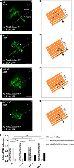
Genetic Loss of Function of Components of the Reelin Pathway Disrupts Lamination of Periventricular Neuron Arbors in the Tectal Neuropil (A and B) Confocal reconstruction (A) and corresponding schematics (B) of a transiently GFP-labeled bistratified PVN in the tectum of a 5 dpf wild-type larva shown from the side parallel to the skin (dotted line) to highlight the PVNs’ laminar morphology. Stratified PVNs did not show any lamination defects. A PVN was classified as displaying lamination defects if we could detect several arbor branches crossing multiple laminae per PVN. (C and D) Confocal reconstruction (C) and corresponding schematics (D) of a transiently GFP-labeled bistratified PVN in the tectum of a 5 dpf reln−/− larva highlighting aberrant PVN morphology. The white arrowheads indicate multiple branches atypically crossing distinct neuropil laminae. (E and F) Confocal reconstruction (E) and corresponding schematics (F) of a transiently GFP-labeled bistratified PVN in the tectum of a 5 dpf vldlr−/− larva showing aberrant PVN morphology. The white arrowheads indicate branches crossing different neuropil laminae. (G and H) Confocal reconstruction (G) and corresponding schematics (H) of a transiently GFP-labeled monostratified PVN in the tectum of a 5 dpf dab1a−/− larva showing aberrant PVN morphology. The white arrowheads indicate branches meandering across different neuropil laminae. (A–H) SO, stratum opticum; SFGS, stratum fibrosum et griseum superficiale; SGC, stratum griseum centrale; SAC, stratum album centrale; D, dorsal; A, anterior. Scale bars, 20 μm. (I) Frequency distribution of non-stratified PVNs, stratified PVNs without, or stratified PVNs with obvious lamination defects in wild-type (n = 14 PVNs from 13 larvae), reln−/− (n = 10 PVNs from 9 larvae), vldlr−/− (n = 9 PVNs from 8 larvae), and dab1a−/− (n = 13 PVNs from 13 larvae) at 5 dpf. Lamination defects of stratified PVNs were defined as the occurrence of several arbor branches originating from the cell body crossing multiple neuropil laminae. PVN frequency distributions were significantly different in reln−/− and dab1a−/− larvae compared to wild-type larvae, respectively (k × 2 chi-square test after Brandt-Snedecor, wild-type versus reln−/− p = 0.0082, wild-type versus dab1a−/− p = 0.0036), whereas there was no significant difference observed between wild-type and vldlr−/− (p = 0.12). These data suggest that genetic loss of function of components of the Reelin pathway also disrupt the ability of PVNs to target single synaptic lamina in the tectum. See also Figure S3. |