
Fig. S5
- ID
- ZDB-FIG-180622-24
- Publication
- Di Donato et al., 2018 - An Attractive Reelin Gradient Establishes Synaptic Lamination in the Vertebrate Visual System
- Other Figures
- All Figure Page
- Back to All Figure Page
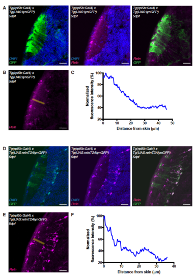
Ectopic expression of reelin in the tectum. Related to Figure 5. (A) Immunostaining of anti-reelin (magenta), anti-GFP (green) and DAPI (blue) on a horizontal cryo-section through the tectum of 5 dpf Tg(rpl5b:Gal4, UAS:lynGFP) transgenic larva expressing membrane tagged lyn-GFP in PVNs and radial glia cells. (B) Anti-reelin staining shown in (A) and the rectangle in yellow along which the Reelin gradient was measured. (C) Densitometric plot of normalized anti-reelin fluorescence intensity taken from tecta expressing GFP indicating that endogenous reelin expression is unaffected by GFP expression (ctrl). (D) Immunostaining of anti-reelin (magenta), anti-GFP (green) and DAPI (blue) on a horizontal cryo-section through the tectum of 5 dpf larva expressing the rpl5b:Gal4; UAS:relnT2AGFP transgenes in PVNs and radial glia cells. Reelin protein is present in PVNs and RG cells expressing the UAS:relnT2AGFP transgene as shown by co-labeling of Reelin and GFP (right panel). (E) Anti-reelin staining shown in (D) and the rectangle in yellow along which the Reelin gradient was measured. (F) Densitometric plot of normalized anti-reelin fluorescence intensity taken from tecta ectopically expressing relnT2AGFP. The overall superficially high to deep low gradient distribution seen in the ctrl seems to be intact however local perturbations of the gradient were observed. Scale bars = 25 μm. |