
Fig. S4
- ID
- ZDB-FIG-180622-23
- Publication
- Di Donato et al., 2018 - An Attractive Reelin Gradient Establishes Synaptic Lamination in the Vertebrate Visual System
- Other Figures
- All Figure Page
- Back to All Figure Page
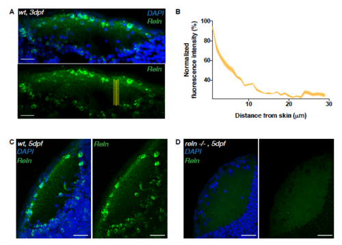
Reelin immunoreactivity in the neuropil of 3 dpf reln-/- larvae. Related to Figure 4. (A) Immunostaining of anti-reelin (green) and DAPI (blue) on a horizontal cryosection of a 3 dpf larval tectum. Scale bar = 20μm. The yellow rectangle indicates the long-axis along which the Reelin gradient in (B) was measured. (B) Densitometric plot of normalized anti-reelin fluorescence intensity taken from 3 different tecta at 3dpf. Data are represented as mean ± s.e.m. (C, D) Horizontal sections of 5dpf tectum showing reelin spatial distribution detected wildtype larva (C) or a reln-/- mutant (D). The reelin protein is absent in the mutant. Scale bars = 30 μm. |
| Gene: | |
|---|---|
| Antibody: | |
| Fish: | |
| Anatomical Term: | |
| Stage Range: | Protruding-mouth to Day 5 |
| Fish: | |
|---|---|
| Observed In: | |
| Stage: | Day 5 |