
Fig. 5
- ID
- ZDB-FIG-180622-18
- Publication
- Di Donato et al., 2018 - An Attractive Reelin Gradient Establishes Synaptic Lamination in the Vertebrate Visual System
- Other Figures
- All Figure Page
- Back to All Figure Page
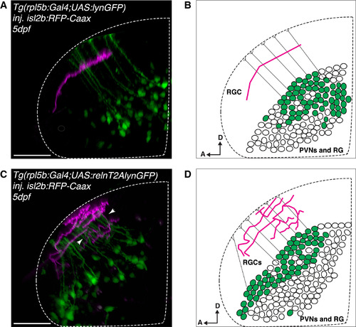
Ectopic Reelin Expression in Periventricular Neurons and Radial Glia Causes Perturbations of Single Laminae Targeting of RGCs in the Tectal Neuropil (A) Lateral projection of a representative confocal stack showing a single RGC axon genetically labeled with isl2b:RFP (magenta) in the tectum of a larva expressing the rpl5b:Gal4;UAS:GFP transgene (control), which is active in a subset of PVNs and radial glia cells (RGs) (green). All analyzed RGCs targeted a single lamina (n = 17/17 fish). Scale bar, 35 μm. (B) Schematics of a single RGC axon targeting a distinct lamina in the tectal neuropil of a 5 dpf larva expressing GFP (control) in a subset of PVNs and RGs (green), thus spatially opposite of the superficial Reelin-enriched neuropil zone (as in A). PVNs, periventricular neurons; RG, radial glia cells; D, dorsal; A, anterior. (C) Lateral projection of a representative confocal stack showing multiple isl2b:RFP-labeled RGC axons (magenta) in the tectum of a Tg(rlp5b:Gal4;UAS:relnT2AlynGFP) larva allowing the co-expression of mouse reelin and membrane-GFP in a subset of PVNs and RGs (green). RGC axons meander between multiple laminae (white arrowheads, altered lamination pattern observed in n = 11/19 larvae), thus suggesting that ectopic reeln expression is interfering with the endogenous graded Reelin distribution. Scale bar, 35 μm. (D) Schematics of multiple RGC axons in the tectal neuropil of a 5 dpf larva expressing relnT2AlynGFP in a subset of PVNs and RGs (green), thus spatially opposite of the superficial Reelin-enriched neuropil zone (as in C). PVNs, periventricular neurons; RGs, radial glia cells; D, dorsal; A, anterior. See also Figure S5. |