
Fig. S10
- ID
- ZDB-FIG-180529-7
- Publication
- Sánchez-Iranzo et al., 2018 - Tbx5a lineage tracing shows cardiomyocyte plasticity during zebrafish heart regeneration
- Other Figures
- All Figure Page
- Back to All Figure Page
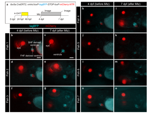
Assessment of efficient ablation of tbx5a-derived cardiomyocytes in individualized fish. a tbx5a+ ventricular cardiomyocytes were genetically ablated in tbx5a:CreERT2;vmhcl:loxP-tagBFP-loxP-mCherry-NTR double transgenic zebrafish by administration of Metronidazole (Mtz). Recombination was induced by administration of 4-Hydroxytamoxifen (4-OHT) at 1 and 2 days postfertilisation (dpf). At 4 dpf, larvae where imaged under a binocular scope. b–o Images show lateral views of the head and heart region. The lens is red (gamma-crystallin transgenic reporter), the cardiac ventricle is red (mCherry+ tbx5a-derived ventricular cardiomyocytes) and blue (tbx5a- cardiomyocytes). Fish were individually treated with Mtz from 4 to 7 dpf. At 7 dpf, 3 days after initiation of Mtz treatment, each larva was imaged again. At this stage, tagBFP expression is visible and expanded but mCherry is no longer detected in most larvae. FHF, first heart field; SHF, second heart field. Scale bars, 100 μm |