
Fig. 10
- ID
- ZDB-FIG-180524-22
- Publication
- Sánchez-Iranzo et al., 2018 - Tbx5a lineage tracing shows cardiomyocyte plasticity during zebrafish heart regeneration
- Other Figures
- All Figure Page
- Back to All Figure Page
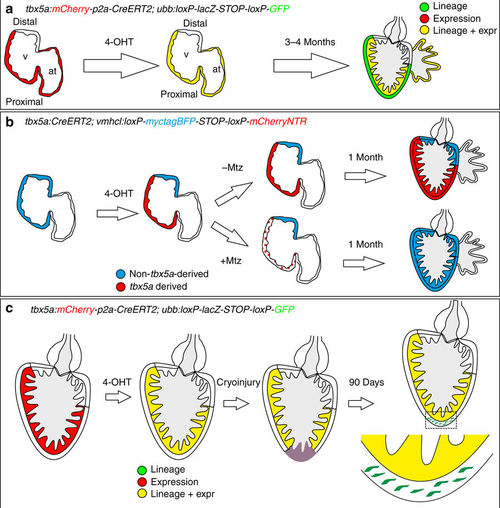
Summary of the contribution of tbx5a-positive and -negative myocardium during heart development and regeneration. a Identification of tbx5a-expressing and tbx5a-derived cells in the zebrafish heart. Red, tbx5a+ cells; green, tbx5a-derived cells not expressing tbx5a; yellow, tbx5a-expressing cells derived from embryonic tbx5a+ cells. b Replacement of the embryonic first heart field myocardium with second heart field progenitors. Blue, ventricular cardiomyocytes; red, tbx5a-derived ventricular cardiomyocytes; dotted red, ablated tbx5a-derived ventricular cardiomyocytes. c Contribution of tbx5a-derived cells during heart regeneration in the zebrafish. Red, tbx5a+ cells; green, tbx5a-derived cells not expressing tbx5a; yellow, tbx5a-expressing cells derived from trabecular adult tbx5a+ cells. 4-OHT, 4-Hydroxytamoxifen; at, atrium; Mtz, Metronidazole; v,ventricle |