
FIGURE
Fig. S5
- ID
- ZDB-FIG-180529-1
- Publication
- Sánchez-Iranzo et al., 2018 - Tbx5a lineage tracing shows cardiomyocyte plasticity during zebrafish heart regeneration
- Other Figures
- All Figure Page
- Back to All Figure Page
Fig. S5
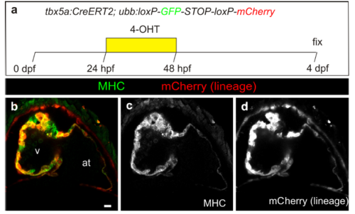
Fate mapping of embryonic tbx5a-derived cells. a–d Optical section through a tbx5a:CreERT2;ubb:loxP-GFP-loxP-mCherry heart at 4 days postfertilisation (dpf) treated with 4-Hydroxytamoxifen (4-OHT) as shown in a revealing efficient recombination in ventricular and atrial cardiomyocytes (mCherry, red). Shown is a section through the atrioventricular canal region. The distal tbx5a- region is not visible in this section. See Supplementary Movie 5 for a full z-stack visualization. at, atrium; hpf, hours postfertilisation; v, ventricle. Scale bars, 10 μm |
Expression Data
Expression Detail
Antibody Labeling
Phenotype Data
Phenotype Detail
Acknowledgments
This image is the copyrighted work of the attributed author or publisher, and
ZFIN has permission only to display this image to its users.
Additional permissions should be obtained from the applicable author or publisher of the image.
Full text @ Nat. Commun.