
FIGURE
Fig. S8
- ID
- ZDB-FIG-180529-5
- Publication
- Sánchez-Iranzo et al., 2018 - Tbx5a lineage tracing shows cardiomyocyte plasticity during zebrafish heart regeneration
- Other Figures
- All Figure Page
- Back to All Figure Page
Fig. S8
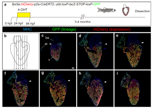
Fate mapping of embryonic tbx5a-derived cells on sagittal sections of adult hearts. a Overview of the experimental setup. b Scheme showing the sectioning orientation through the heart and the location of the individual sections shown in the figure. c–i Immunofluorescence staining of adult heart sections recombined as in a. Shown are merged channels for GFP (green), mCherry (red) and anti-MHC staining (blue). Arrowheads point to the negative basal domain in the ventricle n=3/3. Scale bar, 100 μm |
Expression Data
Expression Detail
Antibody Labeling
Phenotype Data
Phenotype Detail
Acknowledgments
This image is the copyrighted work of the attributed author or publisher, and
ZFIN has permission only to display this image to its users.
Additional permissions should be obtained from the applicable author or publisher of the image.
Full text @ Nat. Commun.