
FIGURE
Fig. S4
- ID
- ZDB-FIG-180208-26
- Publication
- Benjamin et al., 2017 - Intravital imaging of metastasis in adult Zebrafish
- Other Figures
- All Figure Page
- Back to All Figure Page
Fig. S4
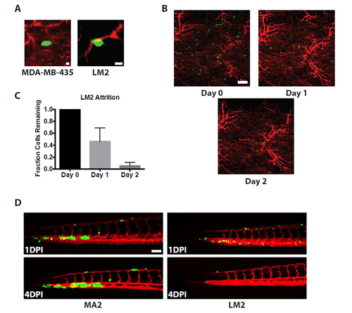
Human tumor cells arrest and extravasate in adult zebrafish but fail to form tumors. (A) Images of human melanoma (MDA-MB-435) and breast cancer (LM2) cells that have extravasated in zebrafish 2 days post-injection. Scale bars are 10 μm. (B) Images showing the attrition of LM2 cells over time in adult zebrafish following injection. Scale bar is 100 μm. (C) Quantification of the fraction of LM2 cells remaining over time in adult zebrafish. n = 47 fields in 7 different fish. (D) Images of the tails of embryos 1 and 4 DPI (3 and 6 days old) injected with LM2 or MA2 cells. Scale bar is 100 μm. |
Expression Data
Expression Detail
Antibody Labeling
Phenotype Data
Phenotype Detail
Acknowledgments
This image is the copyrighted work of the attributed author or publisher, and
ZFIN has permission only to display this image to its users.
Additional permissions should be obtained from the applicable author or publisher of the image.
Full text @ BMC Cancer