
Fig. 1
- ID
- ZDB-FIG-180208-22
- Publication
- Benjamin et al., 2017 - Intravital imaging of metastasis in adult Zebrafish
- Other Figures
- All Figure Page
- Back to All Figure Page
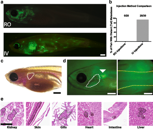
Intravenous injection of zebrafish melanoma cells (ZMEL1) into adult zebrafish. a Representative images of zebrafish injected with GFP-labeled zebrafish ZMEL1 melanoma cells 14 days after retro-orbital (top) and intravenous (bottom) injections. Scale bar is 1 mm. b Quantification of injection efficiency of retro-orbital and intravenous injections as determined by the presence of distant metastases in the posterior of the fish with the success rate indicated. c 6 to 10-week-old casper fish with injection location (common cardinal vein) outlined. Scale bar is 1 mm. d Example of a successful intravenous injection as indicated by GFP-labeled tumor cells in the gills (white dashed line) and posterior of a casper fish (yellow dashed line) 1 h post-injection. The injection site is indicated with a white arrowhead. Scale bar is 1 mm. e H&E stained transverse sections of zebrafish 14 days post-injection showing tumors in the indicated organs. Tumors are indicated by black dotted lines. Scale bar is 100 μm |