
FIGURE
Fig. 2
- ID
- ZDB-FIG-180208-23
- Publication
- Benjamin et al., 2017 - Intravital imaging of metastasis in adult Zebrafish
- Other Figures
- All Figure Page
- Back to All Figure Page
Fig. 2
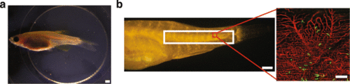
Live imaging of adult zebrafish following injection of ZMEL1 melanoma cells. a Intravital imaging set-up with an adult zebrafish restrained in low-melt agarose in a glass-bottomed 6-well plate. Scale bar is 1 mm. b Region of imaging in the posterior of a casper;flk:dsRed zebrafish (white box) and example of a single 20× confocal field (red box). Scale bar for the posterior is 1 mm. Scale bar for the 20× field is 100 μm |
Expression Data
| Gene: | |
|---|---|
| Fish: | |
| Condition: | |
| Anatomical Term: | |
| Stage: | Adult |
Expression Detail
Antibody Labeling
Phenotype Data
Phenotype Detail
Acknowledgments
This image is the copyrighted work of the attributed author or publisher, and
ZFIN has permission only to display this image to its users.
Additional permissions should be obtained from the applicable author or publisher of the image.
Full text @ BMC Cancer