
Fig. 4
- ID
- ZDB-FIG-180208-25
- Publication
- Benjamin et al., 2017 - Intravital imaging of metastasis in adult Zebrafish
- Other Figures
- All Figure Page
- Back to All Figure Page
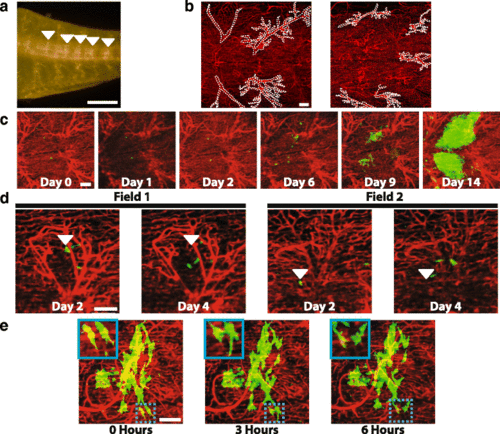
Imaging of disseminated tumor cells over the course of two weeks. a Image of the tail of a flk:dsRed;casper fish with the large vessels used as landmarks indicated by white arrowheads. Scale bar is 1 mm. b Example 10× fields with the large vessels used for landmarks (white dotted lines) highlighted to indicate that the vessels in each field are unique. Scale bar is 100 μm. c One field containing ZMEL1 tumor cells imaged over the course of two weeks showing the growth pattern of metastases. Scale bar is 100 μm. d Images of two sites on day 2 and day 4 post-injection showing that individual cells are rarely found in the same location during this time period. Arrowheads indicate the position of selected cells 2 days post-injection. Scale bar is 100 μm. e One field 9 days post-injection that was imaged over the course of 6 h showing cells extending and retracting protrusions. Inset shows higher magnification of two cells that change shape extensively during the 6 h of imaging. Scale bar is 100 μm |
| Gene: | |
|---|---|
| Fish: | |
| Condition: | |
| Anatomical Term: | |
| Stage: | Adult |