
Fig. 3
- ID
- ZDB-FIG-171228-48
- Publication
- Rességuier et al., 2017 - Specific and Efficient Uptake of Surfactant-Free Poly(Lactic Acid) Nanovaccine Vehicles by Mucosal Dendritic Cells in Adult Zebrafish after Bath Immersion
- Other Figures
- All Figure Page
- Back to All Figure Page
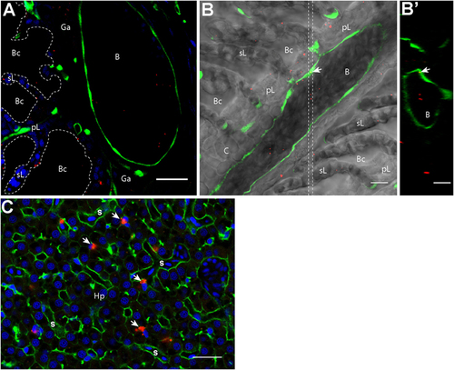
Nanoparticles (NPs) reach the circulatory system and the liver. (A,B′) Representative confocal images of gills from transgenic fli:GFP adults bathed for 24 h in 0.05% red fluorescent NPs. Images were acquired in 40-μm-thick whole-body cryosections, stained with DAPI [blue in (A)]. NPs are found in the lumen of gill arch (A) and gill filament (B) blood vessels, which are delimited by green endothelial cells. NPs were also detected inside endothelial cells [arrow in (B)]. (B′) These observations are confirmed by the orthogonal view realized in the area between the two dotted lines in (B). (C) Representative confocal image of the liver of wild-type adults exposed to 0.01% red fluorescent NPs. Images were acquired in 40-μm-thick whole-body cryosections, stained with phalloidin (green) and DAPI (blue). NPs are highly concentrated in cells close to sinusoids (s) and displaying an oblong nucleus (arrows). pL, primary lamellae; sL, secondary lamellae; Bc, branchial cavity; B, blood vessel; Ga, gill arch; C, cartilage; Hp, hepatocytes. Scale bar: 10 μm (B,B′), 20 μm (A,C). |