
FIGURE
Fig. 2
- ID
- ZDB-FIG-170922-38
- Publication
- Liu et al., 2017 - Zebrafish B Cell Development without a Pre-B Cell Stage, Revealed by CD79 Fluorescence Reporter Transgenes
- Other Figures
- All Figure Page
- Back to All Figure Page
Fig. 2
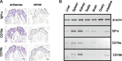
Predominant expression of CD79a and CD79b in kidney by in situ hybridization. (A) In situ hybridization of IgH-μ, CD79a, and CD79b antisense RNA probes to thin sections of zebrafish kidney. Images were processed with Nuance software to highlight hybridization signal stained by NBT/5-bromo-4-chloro-3-indolyphosphate. Right panels show background with sense probes. Scale bars, 400 μm. (B) RT-PCR analysis of CD79a and CD79b in zebrafish tissues. Image color is inverted for clarity. |
Expression Data
| Genes: | |
|---|---|
| Fish: | |
| Anatomical Terms: | |
| Stage: | Adult |
Expression Detail
Antibody Labeling
Phenotype Data
Phenotype Detail
Acknowledgments
This image is the copyrighted work of the attributed author or publisher, and
ZFIN has permission only to display this image to its users.
Additional permissions should be obtained from the applicable author or publisher of the image.
Full text @ J. Immunol.