
Image
|
Figure Caption
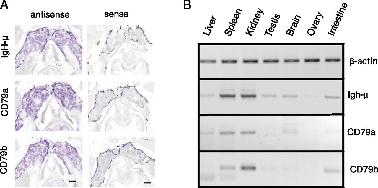
Fig. 2
Predominant expression of CD79a and CD79b in kidney by in situ hybridization. (A) In situ hybridization of IgH-μ, CD79a, and CD79b antisense RNA probes to thin sections of zebrafish kidney. Images were processed with Nuance software to highlight hybridization signal stained by NBT/5-bromo-4-chloro-3-indolyphosphate. Right panels show background with sense probes. Scale bars, 400 μm. (B) RT-PCR analysis of CD79a and CD79b in zebrafish tissues. Image color is inverted for clarity.
Figure Data
Acknowledgments
This image is the copyrighted work of the attributed author or publisher, and
ZFIN has permission only to display this image to its users.
Additional permissions should be obtained from the applicable author or publisher of the image.
Full text @ J. Immunol.