
Fig. 5 S1
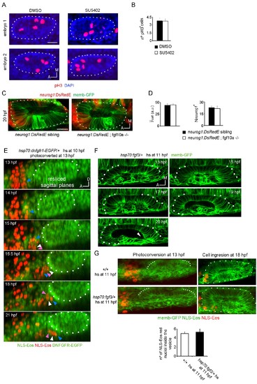
Analysis of cell division controlled by SU5402 and neurog1 expression in FGF10a mutant embryos. (A) pH3 immunostainings at 16 hpf in otic vesicles from DMSO and SU5402 treated embryos. Two embryos in each experimental group are shown. The nuclei were counterstained with DAPI. (B) Quantification of the pH3 immunostainings (n = 12). (C) and (D) neurog1 expression pattern inside the vesicle in neurog1-DsRedE;fgf10a-/- or neurog1-DsRedE; sibling embryos. (C) Z-projection images of vesicles at 20 hpf. (D) Quantifications of the cell and the Nneurog1+ are shown (n = 10 for siblings and n = 5 for fgf10-/-). (E) 3D Tracking of photoconverted NLS-Eos nuclei in hsp70:dnfgfr1-EGFP/+ induced embryos. Z-projections of resliced sagittal sections are shown. Arrowheads indicate examples of tracked nuclei (each color correspond to a different cell). The cells indicated with white and pink arrowheads in the latter panels were not identified in the two first time points, due to their lateral movement out and in of the video during the posterior migration. (F and G) Tissue folding analysis (G) or cell ingression analysis by NLS-Eos photoconversion (F) during placode formation in FGF3 overexpressing embryos (heat-shock at 11 hpf of the Tg(hsp70:fgf3) line). In (F) the posterior region remains unfolded at late stages (20 hpf). Arrowhead: a deformation of the lumen is observed in the posterior region of the vesicle, which is found only anteriorly in wild type embryos associated with the unfolded tissue. Data are mean ± s.e.m. Scale bars, 20 µm. Dotted lines outline the limits of the otic vesicle. |