
Fig. 6
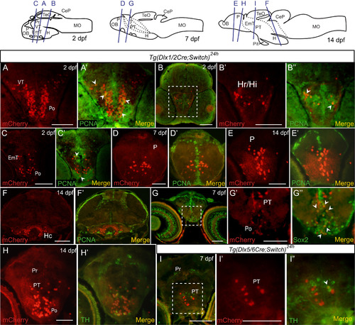
Colocalization of mCherry cells with PCNA, Sox2 and TH in embryos and larvae. (A-I) Double immunohistochemical labeling of cryosections of embryos and larvae. Examples of staining from Tg(dlx1/2Cre;Switch)24h 2 dpf (A-C), 7 dpf (D, G) and 14 dpf (E, F, H) and from Tg(dlx5/6Cre;Switch)24h 7 dpf (I) larvae are shown with relevant brain regions indicated. Each panel has mCherry staining alone (red, left) and merged image with PCNA (A-F), Sox2 (G) and TH (H, I) staining (green, right). The location of each section is depicted in the cartoons at the top. Enlarged versions of the white dotted outlined regions in B, G and I are shown in B'-B'', G'-G'' and I'-I''. Cells that express mCherry and PCNA are indicated with white arrowheads in A', B'', and C', Cells that express mCherry and Sox2 are indicated by white arrowheads in G”. A single cell that expresses mCherry and TH is indicated by a white arrowhead in I''. Scale bar: 100 µm. |
Reprinted from Developmental Biology, 427(1), Solek, C.M., Feng, S., Perin, S., Weinschutz Mendes, H.C., Ekker, M., Lineage tracing of dlx1a/2a and dlx5a/6a expressing cells in the developing zebrafish brain, 131-147, Copyright (2017) with permission from Elsevier. Full text @ Dev. Biol.