
Fig. 5
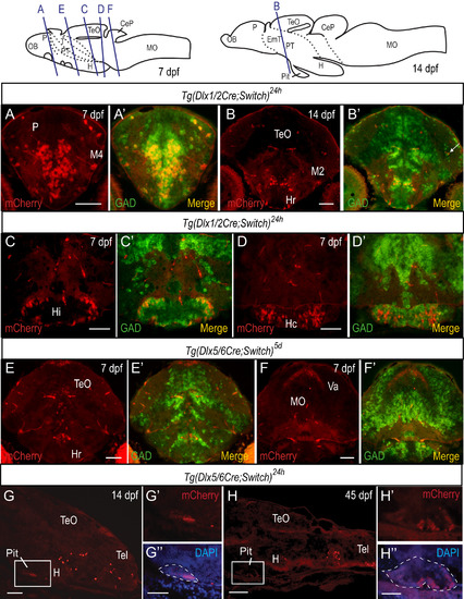
Colocalization of mCherry cells with gad65/67a transcripts in embryos and larvae. (A-F) In situ hybridization (ISH) using gad65 and gad67a probes coupled with immunohistochemistry (IHC) with antibodies against mCherry on cryosections of embryos and larvae. Examples of staining from 7 dpf (A, C-F) and 14 dpf (B) larvae are shown with relevant brain regions indicated. A cell that expresses mCherry but not gad65/67a in the neuropil is indicated by a white arrow in (B’). (G-H) Detection of mCherry protein in sagittal cryosections of Tg(dlx5/6Cre;Switch)24h by immunohistochemistry at 14 dpf (G) and at 45 dpf (juvenile stage, H). Staining is observed in the anterior part of the pituitary, seen best in the higher magnification images (G', G'' and H', H''). DAPI staining (blue) is overlaid with mCherry immunostaining (red) in G” and H”. The pituitary is outlined in white dashed lines. See Table S1 for sample sizes. Scale bars: A-F: 100 µm; G-H: 50 µm; G”, H”: 100 µm. |
Reprinted from Developmental Biology, 427(1), Solek, C.M., Feng, S., Perin, S., Weinschutz Mendes, H.C., Ekker, M., Lineage tracing of dlx1a/2a and dlx5a/6a expressing cells in the developing zebrafish brain, 131-147, Copyright (2017) with permission from Elsevier. Full text @ Dev. Biol.