
Fig. 2
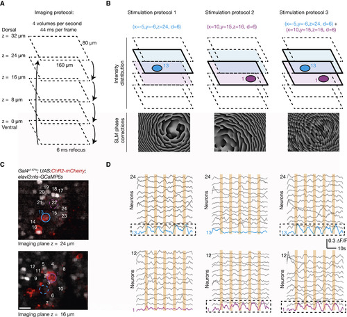
In Vivo 3D High-Resolution Photostimulation and Calcium Imaging (A) Multiplane imaging protocol. An ETL is used to switch rapidly between five imaging planes separated by 8 μm and to record GCaMP6s signals across a volume of 160 × 80 × 32 μm3 at 4 Hz. (B) Design of 3D photostimulation protocols. Based on an acquired z stack, three different 3D photostimulation protocols are designed to target 6-μm stimulation spots either independently or simultaneously to two ChR2-expressing neurons localized in different planes (cell no. 1, purple, in the ventral plane at z = 16 μm; cell no. 13, blue, in the dorsal plane at z = 24 μm). The corresponding phase correction patterns are calculated based on the photostimulation targets and superimposed on the light wavefront by means of the spatial light modulator. (C) A view of the two planes selected for photostimulation from a fish expressing pan-neuronal nuclear localized calcium indicator (nlsGCaMP6s) and ChR2-mCherry. Colored regions of interest (ROIs) indicate the cells selected for the stimulation. The scale bar represents 10 μm. (D) The activity from a subset of cells in the imaged planes during photostimulation. The induced activity matches the spatio-temporal photostimulation protocol (five epochs; 2 s; cell no. 1 and/or cell no. 13), with the targeted cells showing an activity profile locked to the stimulus timing, with minimal cross-activation of the surrounding cells. |